Research · Publication & note
Fabrication and characterization of colloidal crystal thin films - Notes
European Journal of Physics · Journal article · 2 February 2011
Published abstract
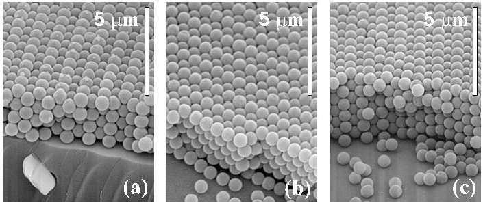
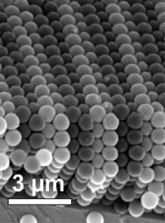
We present a laboratory experiment that allows undergraduate or graduate students to get introduced to colloidal crystal research concepts in an interesting way. Moreover, such experiments and studies can also be useful in the field of crystallography or solid-state physics. The work concerns the growth of colloidal crystal thin films obtained from the crystallization of a latex colloidal solution in a wedge cell. Depending on the thickness of the sample, microcrystals with different structures and orientation are obtained. Colloidal arrangements are studied by scanning electronic microscopy images of the top and edge views of several areas of the crystals.
Figures
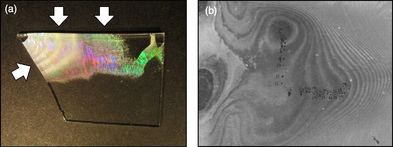
From I. Rodríguez et al., European Journal of Physics 32, 505-515 (2011), DOI 10.1088/0143-0807/32/2/021. © 2011 IOP Publishing Ltd. Reused by the authors under IOP author rights.
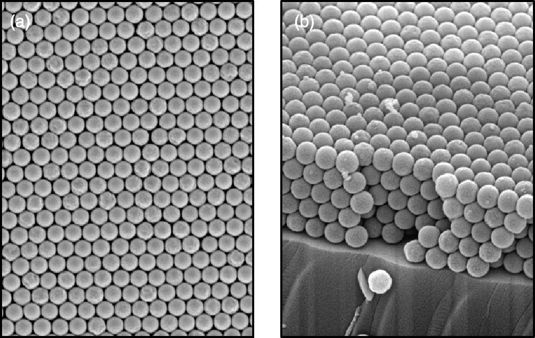
From I. Rodríguez et al., European Journal of Physics 32, 505-515 (2011), DOI 10.1088/0143-0807/32/2/021. © 2011 IOP Publishing Ltd. Reused by the authors under IOP author rights.
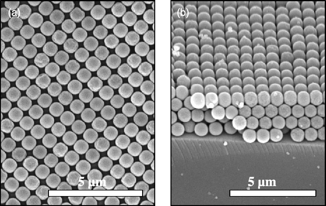
From I. Rodríguez et al., European Journal of Physics 32, 505-515 (2011), DOI 10.1088/0143-0807/32/2/021. © 2011 IOP Publishing Ltd. Reused by the authors under IOP author rights.
How to cite
Article access and reuse
This article was published on a subscription basis. IOP Publishing allows named authors to reuse their original figures with adequate citation and copyright notice, and permits posting of the Accepted Manuscript subject to the applicable author-rights terms and provenance statement. The local file reviewed for this catalogue is the typeset Version of Record, so the PDF is not hosted here. Three complete original figures are reproduced below with full credit. Use the DOI or publisher link to access the journal record.
Related publications

Research fields
Bottom-up
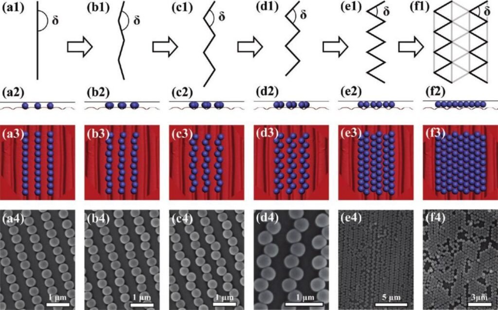
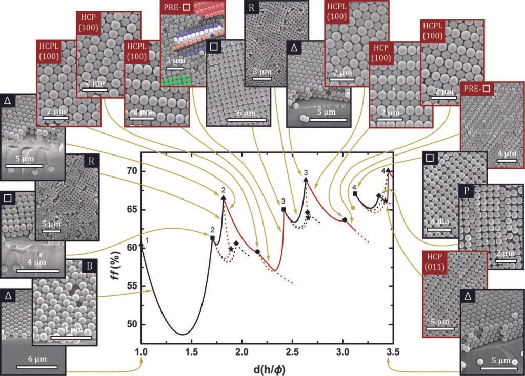
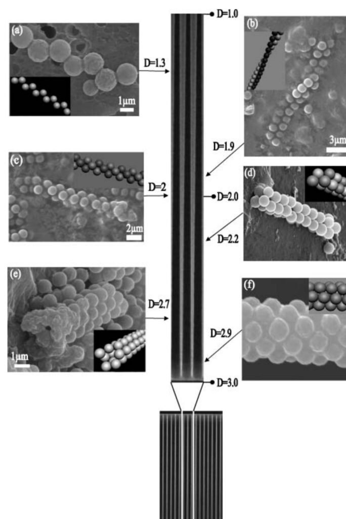
Latex spheres crystallise from suspension inside a wedge cell, where the continuously changing gap selects different layer numbers, orientations and transition structures. Drying converts this confined self-assembly process into an accessible thin-film model of crystal growth.
Simulations & fits
Geometrical FCC and HCP packing models translate top and edge views into three-dimensional particle arrangements. They provide a structural reference for identifying orientations and intermediate phases from microscopy rather than relying on surface symmetry alone.
Characterization
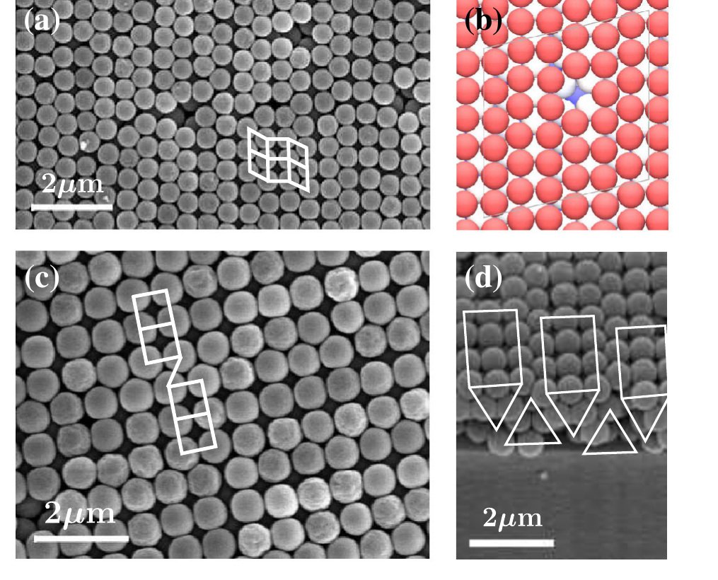
Top-view and cleaved-edge SEM images are acquired along the wedge and compared with optical inspection. The paired perspectives let the experiment distinguish stacking, facet orientation and layer number across a single colloidal film.
RESEARCH TOPICS
 MaterialsTop and cross-sectional SEM images distinguish FCC structures viewed along (111) and (100), as well as an HCP (100) arrangement. The experiment makes confinement visible at particle scale: identical spheres adopt different lattices solely because the local cell thickness changes.
 SetupsThe protocol is deliberately reproducible as an advanced teaching experiment. A Jeol JSM-6300 SEM records both top and edge views; those micrographs are then compared with interactive three-dimensional models, so students connect a two-dimensional image with the underlying stacking sequence.