Research · Publication & note
Layering transitions in confined colloidal crystals: The hcp-like phase - Notes
Rapid Communication
Published abstract
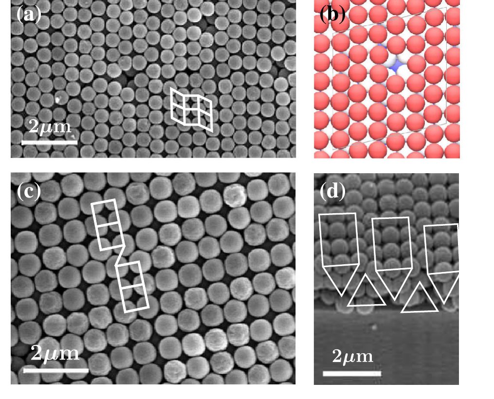
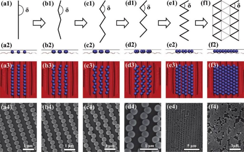
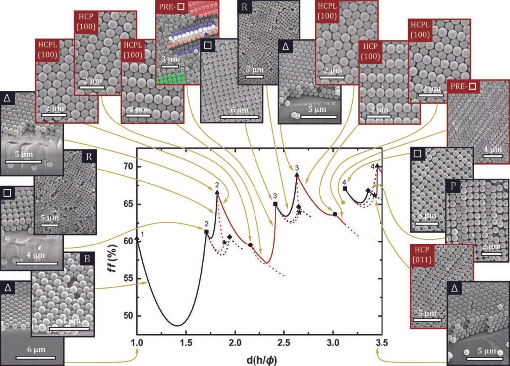
This paper investigates the sequence of morphological transitions in a nearly hard sphere arrangement confined in a wedge cell. A model that shows smooth transitions between the different particle orderings for a small range of confinement is proposed. The hcp-like phase appears as a general adaptive structure in the transitions between two and six layers.
Figures
From F. Ramiro-Manzano et al., Phys. Rev. E 76, 050401(R) (2007), DOI 10.1103/PhysRevE.76.050401. Reproduced under APS author rights.
From F. Ramiro-Manzano et al., Phys. Rev. E 76, 050401(R) (2007), DOI 10.1103/PhysRevE.76.050401. Reproduced under APS author rights.
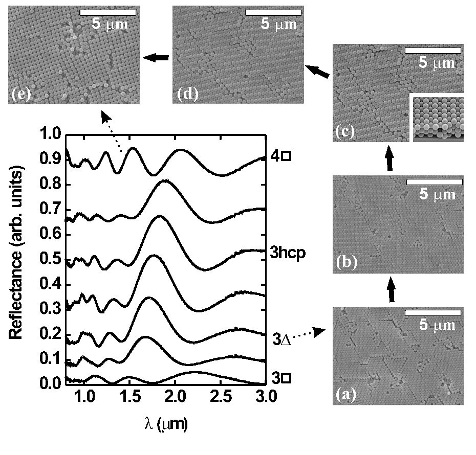
From F. Ramiro-Manzano et al., Phys. Rev. E 76, 050401(R) (2007), DOI 10.1103/PhysRevE.76.050401. Reproduced under APS author rights.
From F. Ramiro-Manzano et al., Phys. Rev. E 76, 050401(R) (2007), DOI 10.1103/PhysRevE.76.050401. Reproduced under APS author rights.
How to cite
Article access and reuse
APS author rights permit authors to use all or part of an APS-published article, including APS-prepared figures, on the author's or employer's website without charging a fee. The four complete figures below were extracted from the local PDF at 300 dpi and are reproduced without modification.
Related publications

Research fields
Bottom-up
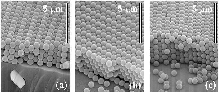
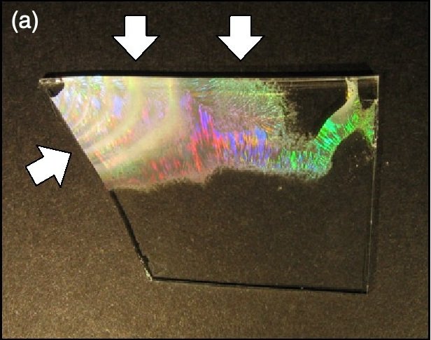
Spheres confined in a wedge cell assemble into layer-dependent colloidal crystals as the plate separation changes. Between conventional triangular and square packings they form an hcp-like arrangement that continuously adapts its distortion to the available gap.
Simulations & fits
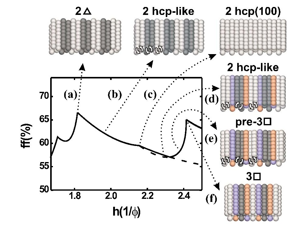
Filling-fraction calculations model buckling and hcp(100) order as limiting cases of one continuous family. This provides a geometrical pathway through the transition and predicts where each intermediate arrangement should be stable.
Characterization
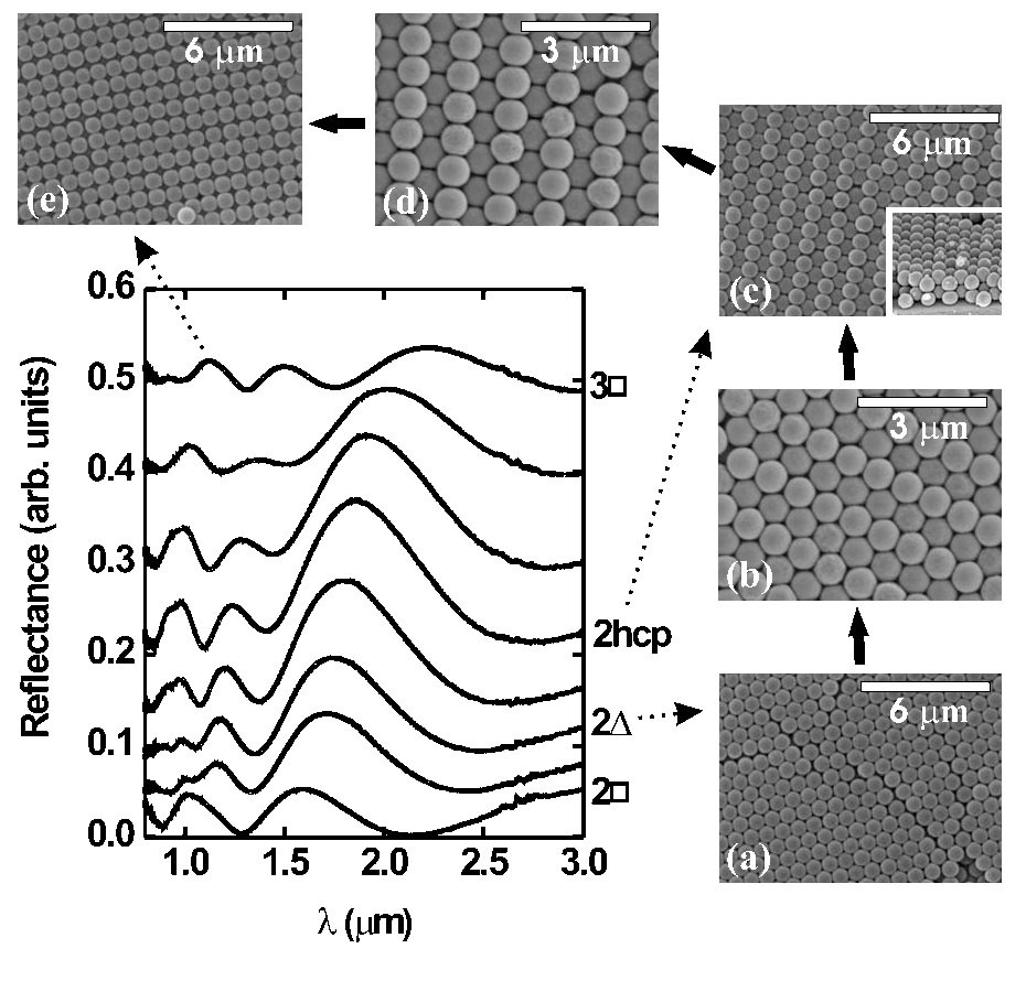
Position-resolved infrared reflectance and SEM imaging follow the same thickness gradient. Spectral discontinuities locate phase boundaries, while surface and edge micrographs identify the particle stacking responsible for each optical change.