Research · Publication & note
Porous silicon microspheres: synthesis, characterization and application to photonic microcavities - Notes
Journal of Materials Chemistry · Journal article · 14 May 2010
Published abstract
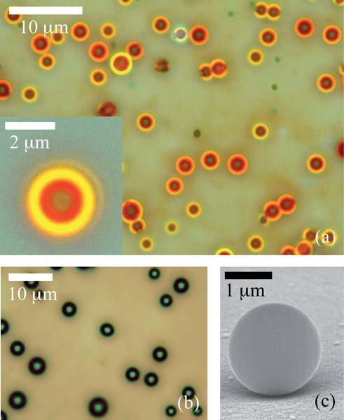
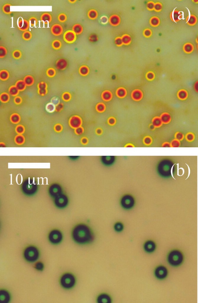
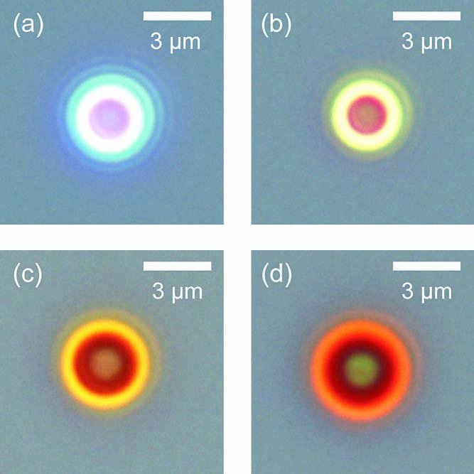
Porous silicon microspheres have been synthesized by chemical vapour deposition of disilane gas. Their spectral signatures are similar to those of electrochemically grown porous silicon, in particular they yield photoluminescence and they show an oxidation behaviour upon their exposure to the open air. The particles are highly spherical and poly-disperse in size with diameters of approximately 0.5 to 5 micrometres, and they have a very smooth surface. Because of these reasons they work as optical microcavities with well defined resonating Mie modes. These modes have been identified in the near-infrared range. They blue-shift considerably whenever the microspheres are in contact with air because of the aforementioned oxidation process.
Figures
From R. Fenollosa et al., J. Mater. Chem. 20, 5210-5214 (2010), DOI 10.1039/C0JM00079E. © The Royal Society of Chemistry 2010. Reproduced by an author with acknowledgement.
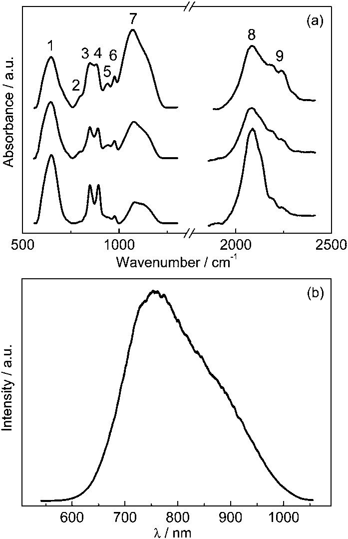
From R. Fenollosa et al., J. Mater. Chem. 20, 5210-5214 (2010), DOI 10.1039/C0JM00079E. © The Royal Society of Chemistry 2010. Reproduced by an author with acknowledgement.
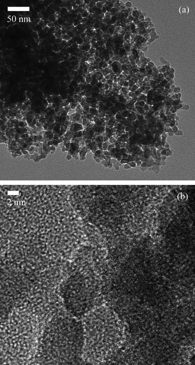
From R. Fenollosa et al., J. Mater. Chem. 20, 5210-5214 (2010), DOI 10.1039/C0JM00079E. © The Royal Society of Chemistry 2010. Reproduced by an author with acknowledgement.
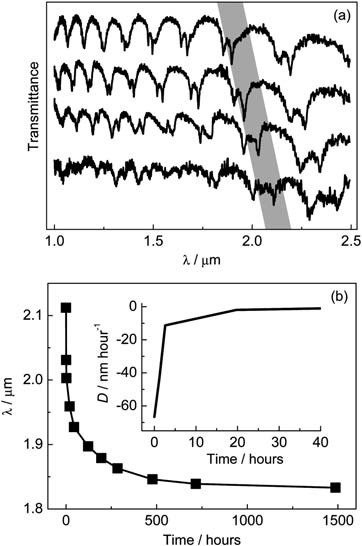
From R. Fenollosa et al., J. Mater. Chem. 20, 5210-5214 (2010), DOI 10.1039/C0JM00079E. © The Royal Society of Chemistry 2010. Reproduced by an author with acknowledgement.
How to cite
Article access and reuse
The subscription Version of Record is not hosted here. RSC states that an author may reproduce figures and diagrams from their own article without requesting permission when correct acknowledgement is given. The figures below are the original compositions extracted from the article PDF and carry full source acknowledgement.
Related publications

Research fields
Bottom-up
Controlled disilane decomposition forms micrometre-scale porous silicon spheres with smooth surfaces and intrinsic photoluminescence. Processing time and subsequent oxidation tune porosity and composition while the spherical boundary supplies cavity confinement.
Simulations & fits
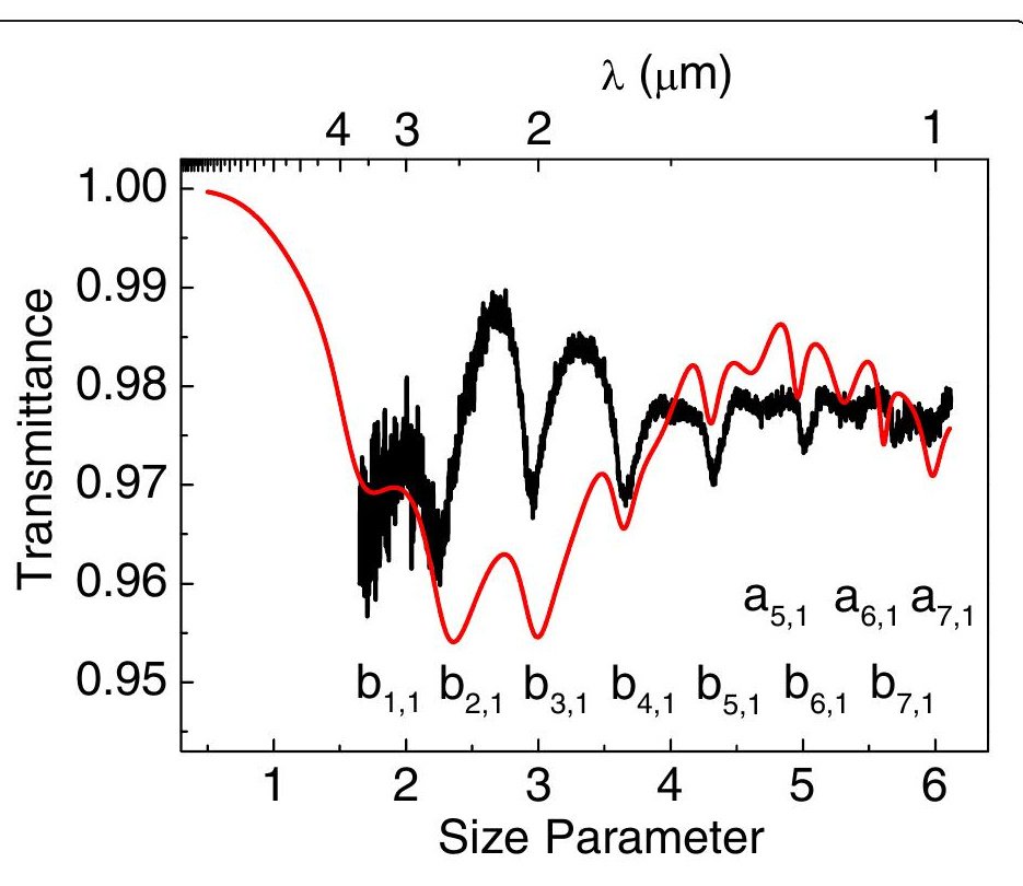
Mie-mode analysis assigns the resonances in single-particle transmission and relates their spectral displacement to effective refractive-index changes. This turns oxidation-induced blue shifts into a sensitive probe of the evolving porous sphere.
Characterization
Optical and electron microscopy, mid-infrared absorption, transmission and photoluminescence characterize morphology, silicon-hydride chemistry and cavity response. Correlating these measurements shows how an emitting porous material and a resonator coexist in one particle.
RESEARCH TOPICS
 EmissionFigure 2 compares their mid-infrared signatures and PL, establishing that they share optical characteristics with electrochemically etched porous silicon despite being free spherical particles. This simultaneous scattering and emission makes the material self-reporting: no separate dye is needed to see the colloids or to interrogate their internal optical evolution.
 MaterialsShort disilane decomposition times of about 1 min 30-40 s produce highly porous, polydisperse silicon spheres. TEM and HRTEM in Figure 3 reveal a polycrystalline skeleton, although a rapidly formed amorphous oxide layer complicates direct lattice interpretation.
 ResonatorsNear-infrared transmission reveals well-defined Mie modes, demonstrating that even a porous and internally heterogeneous sphere can act as a photonic microcavity. The resonance positions therefore provide a sensitive, non-destructive readout of ageing inside the porous silicon framework.