Research · Publication & note
Porous Silicon Microcavities Based Photonic Barcodes - Notes
Advanced Materials · Journal article · 2 May 2011
Article summary
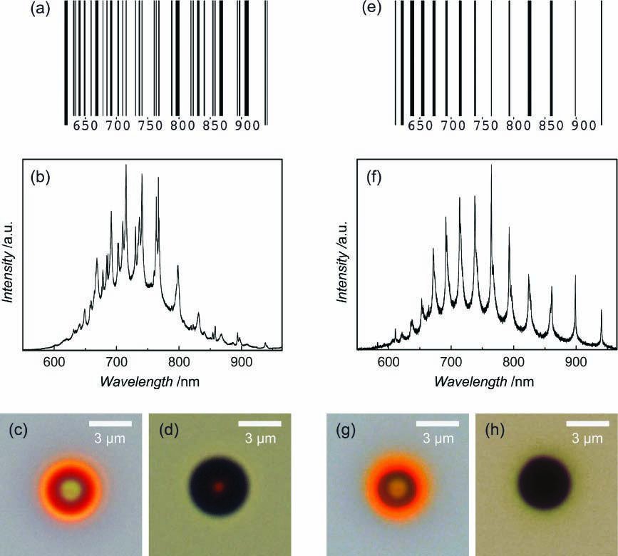
Here, we show that the photoluminescence emission of a single porous silicon colloid is strongly coupled to the whispering-gallery modes of the microcavity defined by the colloid itself, resulting in a unique photoluminescence spectrum with a high quantum yield. This spectrum depends on the colloid size and porosity, and constitutes a fingerprint of the particle. The authors propose a photonic encoding procedure in which a unique barcode can be assigned to each colloid through its photoluminescence spectrum.
Figures
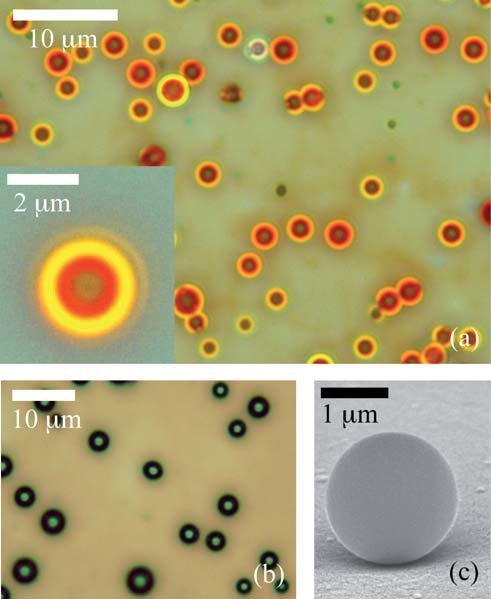
From F. Ramiro-Manzano et al., Advanced Materials 23, 3022-3025 (2011), DOI 10.1002/adma.201100986. © 2011 Wiley-VCH Verlag GmbH & Co. KGaA. Reused by the authors under Wiley's contributor reuse terms.
From F. Ramiro-Manzano et al., Advanced Materials 23, 3022-3025 (2011), DOI 10.1002/adma.201100986. © 2011 Wiley-VCH Verlag GmbH & Co. KGaA. Reused by the authors under Wiley's contributor reuse terms.
From F. Ramiro-Manzano et al., Advanced Materials 23, 3022-3025 (2011), DOI 10.1002/adma.201100986. © 2011 Wiley-VCH Verlag GmbH & Co. KGaA. Reused by the authors under Wiley's contributor reuse terms.
How to cite
Article access and reuse
Wiley permits authors to reuse the article abstract, figures, tables, artwork and selected text within its stated limits, with full credit and without modification. The subscription Version of Record is not hosted here because Wiley's sharing policy restricts public posting of the publisher PDF. Use the DOI or publisher link to access the journal record.
Related publications
Research fields
Bottom-up
Porous silicon microspheres are produced as luminescent, high-index colloids with a radial pore distribution and smooth outer boundary. Each particle therefore forms its own microcavity rather than requiring a patterned mirror stack.
Simulations & fits
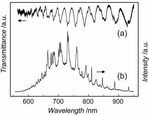
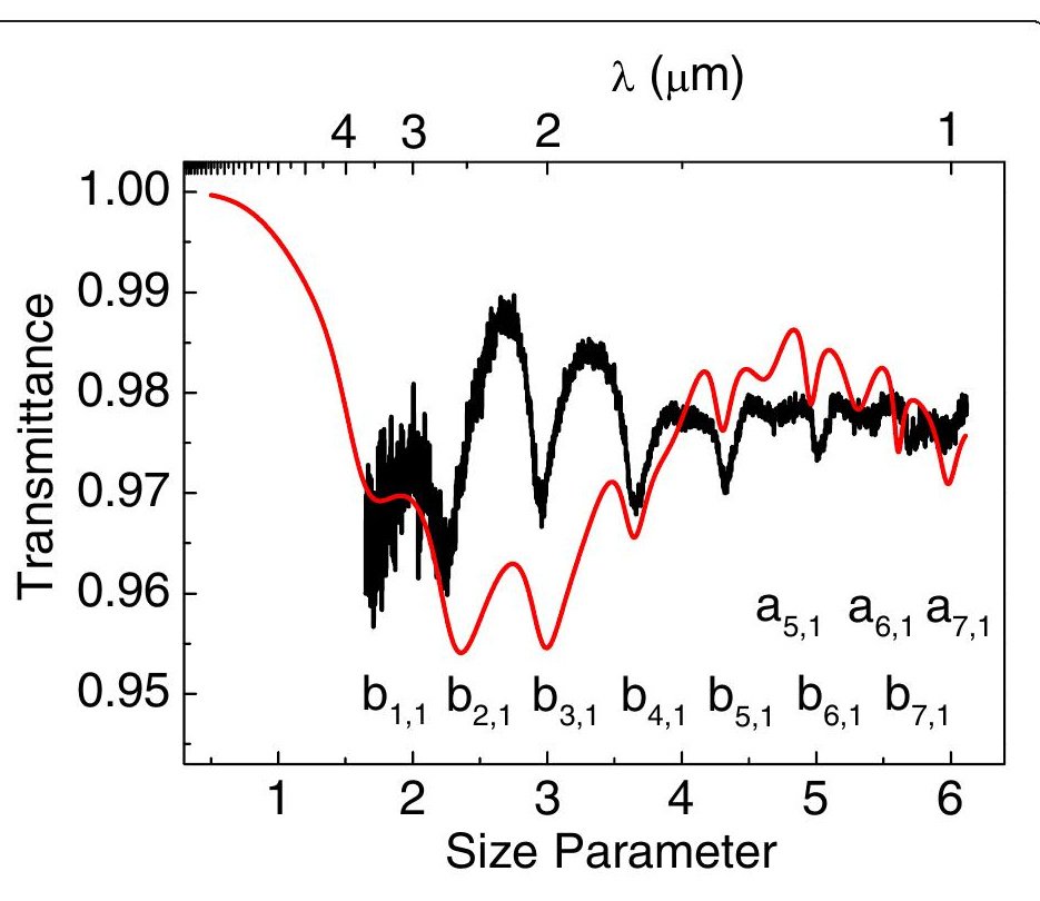
Mie-mode assignments link transmission minima to the resonances that enhance the broad porous-silicon photoluminescence. The resulting sequence of narrow emission peaks constitutes a particle-specific optical barcode governed by diameter and effective refractive index.
Characterization
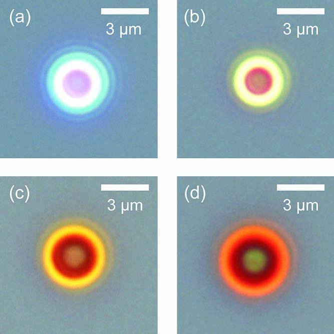
Single-particle transmission and photoluminescence are correlated with optical microscopy and compositional analysis. Measuring the same sphere in both channels shows directly that its luminescence maxima coincide with cavity modes rather than with unrelated spectral structure.
RESEARCH TOPICS
 EmissionMeasuring both channels on the same micromanipulated sphere distinguishes microcavity enhancement from a chemical change in the emitter. The narrow spectral pattern provides a possible optical code readable through luminescence or elastic scattering.
 InterferenceThe barcode is created by the wavelength-dependent constructive and destructive circulation of light inside a sphere. The work uses that reproducible resonance pattern as information, analogous to a spectral barcode, instead of adding multiple dyes with overlapping broad bands.
 MaterialsTheir porosity lowers the effective index and allows biological media to enter the structure, while the silicon skeleton supplies emission. The combination of size, porosity and intrinsic signal is what makes them candidates for encoded labels.
 ResonatorsEach sphere is a three-dimensional Mie cavity. Rather than relying on one colour, the proposed encoding uses several resonance positions, increasing the number of distinguishable labels while keeping the particle micrometre-sized.
 ChemistryPorous silicon provides a chemically modifiable surface without introducing a separate organic luminophore. Because the code depends on cavity geometry as well as luminescence, surface functionalisation can in principle be used for bioconjugation while leaving the resonance sequence readable.